
Ongoing efforts to improve the resolution and robustness of imaging of the pancreas using. Compared to the other techniques magnetic resonance imaging MRI has irreplaceable advantages and can provide valuable.

Endoscopic Ultrasound For Detecting Small Pancreatic Tumor Missed By Computed Tomography Sciencedirect
Magnetic resonance imaging MRI can be used in imaging for PaCa in patients with equivocal findings at ultrasound or MDCT.

Cancer pancreas magnetic resonance imaging. Authors Regina G H Beets-Tan 1 Doenja M J Lambregts 2 Monique Maas 1 Shandra Bipat 3 Brunella Barbaro 4. A comprehensive study design from the consortium for the study of chronic pancreatitis diabetes and pancreatic cancer. Magnetic resonance imaging MRI is a valuable tool in the assessment of the full spectrum of pancreatic disease.
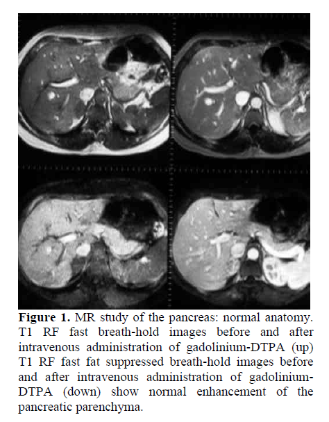
A standard MR protocol including noncontrast T1-weighted fat-suppressed and dynamic gadolinium-enhanced gradient-echo imagings is sensitive for the evaluation of pancreatic cancer. Magnetic resonance cholangiopancreatography MRCP is a special type of MRI that uses computer software to image the pancreatic and bile ducts areas where tumors often form. Gadolinium GdDTPAenhanced T1 and T2weighted standard sequences.
12 However a proper detection and characterization of pancreatic lesions is mandatory because the prognosis is linked to tumor type and grade 3 and correct staging. Epub 2017 Oct 17. MRI examination of the pancreas is done with intravenous administration of contrast material and gadolinium is the most commonly used agent.
Diagnosis of pancreatic cancer remains challenging due to overlapping imaging features with benign lesions notwithstanding great advances with multidetector computed tomography and magnetic resonance imaging MRI. A standard MR protocol including noncontrast T1-weighted fat-suppressed and dynamic gadolinium-enhanced gradient-echo imagings is sensitive for the evaluation of pancreatic cancer. Cancers EISSN 2072-6694 Published by MDPI Disclaimer The statements opinions and data contained in the journal Cancers are solely those of the individual authors and contributors and not of the publisher and the editors.
Magnetic resonance imaging as a non-invasive method for the assessment of pancreatic fibrosis MINIMAP. For adenocarcinoma which is the most common malignant pancreatic tumor helical CT has been most often used for detection and staging but it has limitations in the detection of small. The aim of this study was to determine the diagnostic accuracy of expanded criteria in nodal staging in PDAC patients.
Magnetic resonance imaging MRI is a valuable tool in the assessment of the full spectrum of pancreatic disease. High-resolution HR magnetic resonance imaging MRI has become an indispensable tool for multidisciplinary teams MDTs addressing rectal cancer. MRCP is also used to see pancreatic cysts and blockages in the ducts.
MDPI stays neutral with regard to jurisdictional claims in published maps and institutional affiliations. Further Information Article Processing. Magnetic resonance imaging MRI is a reliable and accurate imaging method for the evaluation of patients with pancreatic ductal adenocarcinoma PDAC.
Its superior soft tissue contrast is beneficial for detection of small non-contour deforming tumors and for detection of a minor portion of PDACs that are isoattenuating on CT. Pancreatic cancer is one of the most common malignant tumors and remains a treatment-refractory cancer with a poor prognosis. Updated recommendations from the 2016 European Society of Gastrointestinal and Abdominal Radiology ESGAR consensus meeting Eur Radiol.
The high mortality associated with pancreatic adenocarcinomas has spurred much interest in developing effective screening tools with MRI using magnetic resonance cholangiopancreatography MRCP playing a central role in the hopes of identifying cancers at earlier stages amenable to curative resection. In our study we report a magnetic resonance imaging MRI approach to visualize the pancreas in mice and to monitor orthotopically implanted pancreatic tumors. Over the last 2 decades computed tomography has become the dominant imaging modality for both the diagnosis and staging of pancreatic cancer with advances in multidetector computed tomography technology improving the ability to identify small tumors and demonstrate subtle degrees of vascular involvement by tumor.
And 2 applications of artificial intelligence. Currently the diagnosis of pancreatic neoplasm depends mainly on imaging and which methods are conducive to detecting small lesions. Technical advances of magnetic resonance imaging MRI including ultrahigh-field magnetic resonance at 30 T parallel imaging techniques and multichannel receive coils of the abdomen have promoted MRI of the pancreas.
However magnetic resonance imaging and positron emission tomography are. Ongoing efforts to improve the resolution and robustness of imaging of the pancreas using. Diffusion-weighted imaging DWI is a relatively recent technological improvement that expanded MRI capabilities having brought functional aspects into conventional morphologic MRI evaluation.
Lymph node staging of ductal adenocarcinoma of the pancreatic head PDAC by cross-sectional imaging is limited. An MRI scanner was used to image normal murine pancreas and the pancreas of mice implanted with a human pancreatic adenocarcinoma cell line. Magnetic resonance imaging as a non-invasive method for the assessment of pancreatic fibrosis MINIMAP.
Magnetic resonance imaging for clinical management of rectal cancer. One of the objectives of the MDT is the preoperative identification of high. The high mortality associated with pancreatic adenocarcinomas has spurred much interest in developing effective screening tools with MRI using magnetic resonance cholangiopancreatography MRCP playing a central role in the hopes of identifying cancers at earlier stages amenable to curative resection.
Optimal use of MRI in the investigation of pancreatic. A comprehensive study design from the consortium for the study of chronic pancreatitis diabetes and pancreatic cancer. To investigate the microcirculation in pancreatic cancer by pharmacokinetic analysis of multiple breath-hold dynamic contrast-enhanced magnetic resonance imaging at 30T.
Quantitative imaging offers several advantages including increased reproducibility and sensitivity to detect mild or diffuse disease. In the past few years there have been two major developments with potential for a significant impact in establishing imaging biomarkers for PDAC and pancreatic cancer premalignancy. Magnetic resonance imaging MRI is often seen as a problem-solving tool for detection and characterization of PDACs 10.
DWI can depict the random. It provides anatomic information for surgical planning and allows patients to be stratified into different groups according to the risk of local and distant recurrence. 1 hyperpolarized metabolic HP-magnetic resonance MR which increases the sensitivity of conventional MR by over 10000-fold enabling real-time metabolic measurements.
Multiple breath-hold dynamic contrast-enhanced magnetic resonance imaging was performed in 40 healthy volunteers and 40 patients with pancreatic cancer proven by histopathology using an. Sixty-six patients with histologically confirmed PDAC that underwent primary surgery were included in this retrospective IRB-approved study. The role of multiparametric mapping MRI in characterizing various tissue types in pancreatic disease such as chronic pancreatitis CP and pancreatic ductal.
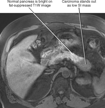
A MRCP can happen at the same time as an MRI. PaCa is hypointense on gadolinium-enhanced T1-weighted images in the pancreatic and venous phases. BackgroundCurrent magnetic resonance imaging MRI of pancreatic disease is qualitative in nature.

Magnetic Resonance Imaging Mri Scan Of The Abdomen Demonstrating A Download Scientific Diagram

Magnetic Resonance Imaging Of Less Common Pancreatic Malignancies And Pancreatic Tumors With Malignant Potential European Journal Of Radiology Open

Normal Pancreatic Appearance On Magnetic Resonance Imaging A Axial Download Scientific Diagram
Pet Mri Ct Metrics Assess Pathologic Response Of Pancreas Cancer To Neoadjuvant Therapy Imaging Technology News

Episode 1 Primary Pancreatic Tumor Diffuse Pancreatic Mass In A Download Scientific Diagram
Challenges In Diagnosis Of Pancreatic Cancer
Challenges In Diagnosis Of Pancreatic Cancer

Diffusion Weighted Mr Imaging Of Pancreatic Cancer A Comparison Of Mono Exponential Bi Exponential And Non Gaussian Kurtosis Models European Journal Of Radiology Open
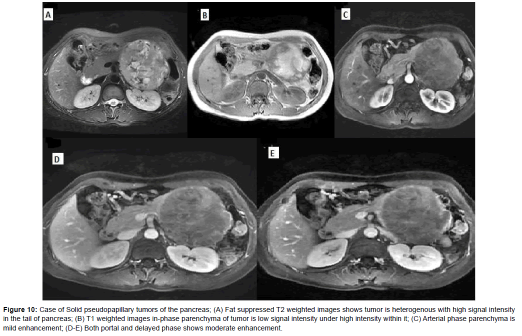
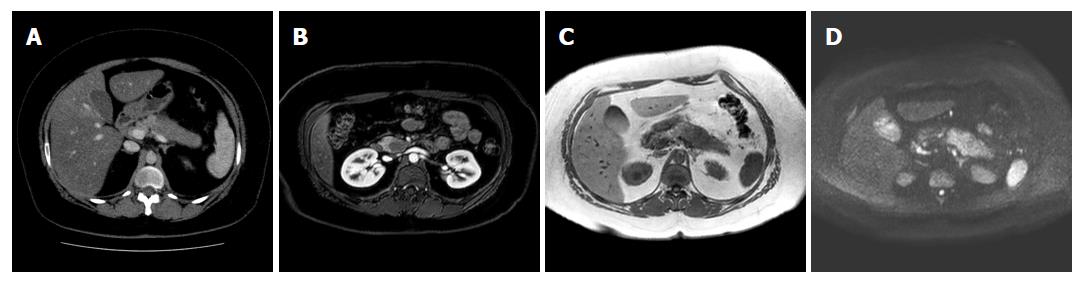
The Value Of Mri In Diagnosis Of Solid Pancreatic Tumors

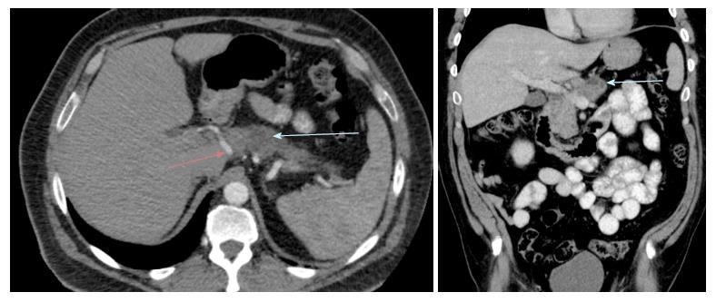
A C Pancreatic Magnetic Resonance Imaging Showed A 2 2 Cm Relatively Download Scientific Diagram

Mri Images Of Histological Proven Pancreatic Cancer In 64 Year Old Man Download Scientific Diagram
Pancreas Radiology Key

Imaging Diagnosis Of Pancreatic Cancer Ct And Mri Oncohema Key

Magnetic Resonance Imaging Of Less Common Pancreatic Malignancies And Pancreatic Tumors With Malignant Potential European Journal Of Radiology Open

Early Diagnosis Of Pancreatic Cancer Current Trends And Concerns Hanada 2017 Annals Of Gastroenterological Surgery Wiley Online Library
Frontiers Magnetic Resonance Guided Radiation Therapy For Pancreatic Adenocarcinoma Advantages Challenges Current Approaches And Future Directions Oncology

View Image
Pancreatic Cancer Mri Protocol For High Risk Patients Healthmanagement Org
Pancreatic Head Mass What Can Be Done Diagnosis Magnetic Resonance Imaging Insight Medical Publishing


Komentar